We build biophysical models and imaging tools that turn diffusion MRI into quantitative markers of brain tissue — and translate them from an ultra-strong-gradient research scanner to the clinic

RICE: orientation-independent biomarkers validated across a 1000+ subject MS cohort
Axonal shape parameters in seconds instead of months of simulation
Biophysical axonal markers across development, stroke, and multiple sclerosis (N = 821)
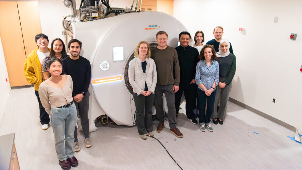
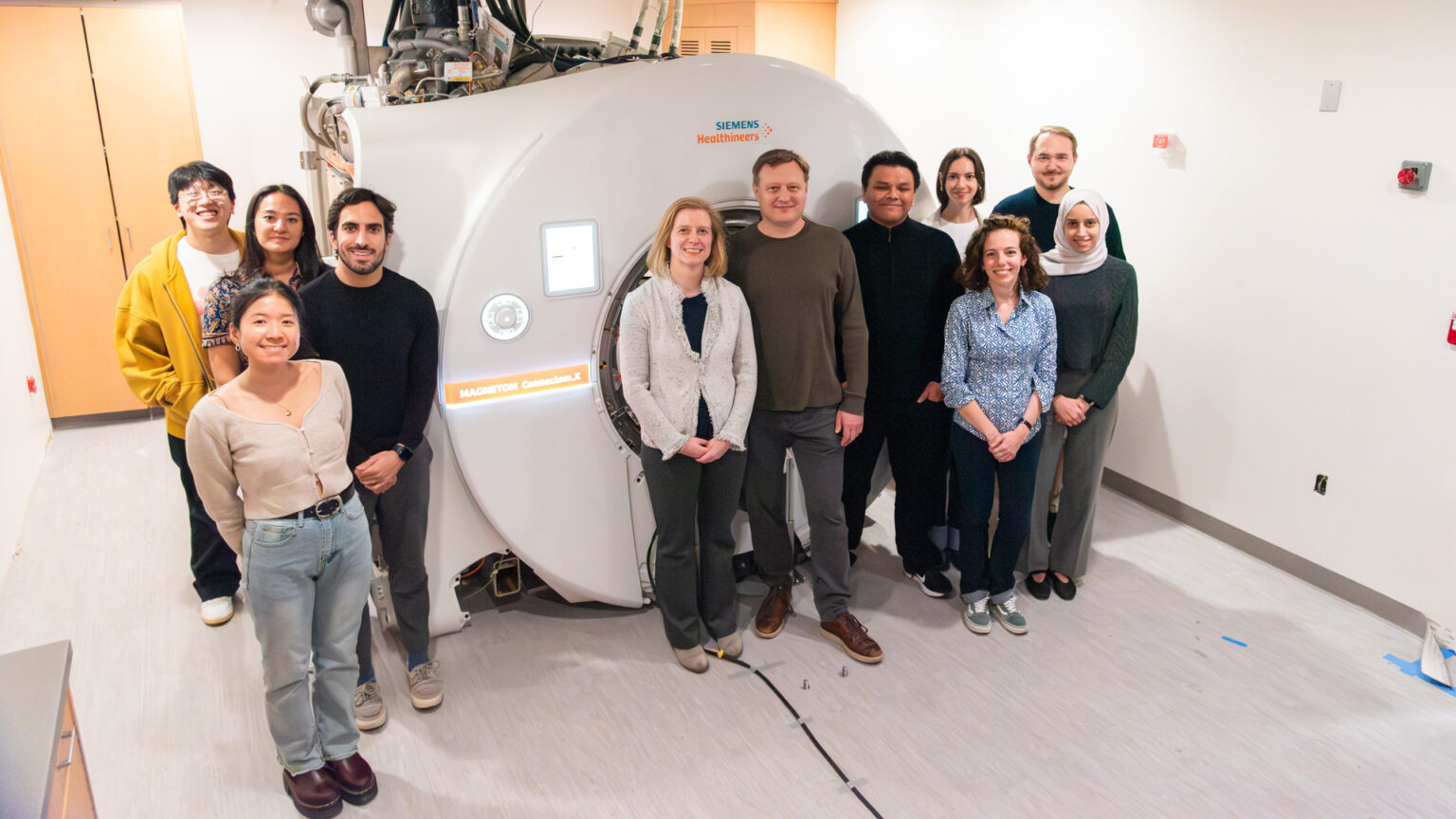
We are the MRI Biophysics Group at NYU Grossman School of Medicine, in the Center for Biomedical Imaging, co-directed by Els Fieremans and Dmitry Novikov. Our work spans the physics of diffusion in tissue, the algorithms that estimate microstructure from the MRI signal, and their translation into clinical markers for neurodegeneration, MS, traumatic brain injury, and aging.
Meet the team →
Diffusion is not a single number — it changes with the timescale over which you observe it, and that time-dependence D(t) encodes the structural universality class of the tissue: how barriers and restrictions are arranged. Measuring how D(t) approaches its long-time limit reveals cell sizes, membrane permeability, surface-to-volume ratio — and, via scattering theory, the structural disorder along and across axons.
Novikov et al., Nature Physics 2011 · Novikov et al., PNAS 2014 · Fieremans et al., NeuroImage 2016 · Lee et al., Communications Biology 2020 · Abdollahzadeh et al., Nature Communications 2025
The diffusion signal carries a faint imprint of axon size — weighted toward the largest fibers and floored by the smallest, so the measured quantity is an effective radius reff, not a simple mean. We established how to recover it from the 1/√b signal scaling at high diffusion weighting, and how realistic axon shape — beading, undulations — biases it.
Veraart et al., eLife 2020 · Lee et al., NeuroImage 2020 · Lee et al., NMR in Biomedicine 2024
White matter behaves, to the diffusion signal, as multiple compartments — water inside narrow axons and water around them — sharing one fiber-orientation distribution. The Standard Model unifies WMTI, NODDI, ball-and-stick, Fiber Ball and other models of multiple Gaussian compartments as special cases of one framework, with reproducible clinical estimation and histological validation.
Novikov et al., NeuroImage 2018 · Coelho et al., NeuroImage 2022 · Coronado-Leija et al., Imaging Neuroscience 2024
In gray matter, water crosses neuronal membranes fast enough to carry information standard diffusion metrics cannot see. NEXI adds a fourth parameter — the exchange time τex (≈ 13 ms in human cortex). Because exchange this fast needs short diffusion times at high gradient strength, NEXI is the primary scientific rationale for the Connectom.X.
Jelescu et al., NeuroImage 2022 · Chan et al., Imaging Neuroscience 2025
Our biophysical markers translate from theory to the clinic — turning diffusion MRI into quantitative, interpretable biomarkers of disease: demyelination and axonal loss in multiple sclerosis, gray- and white-matter change in Alzheimer's disease and aging, and tissue injury in stroke and traumatic brain injury.
Diffusion MRI is, at root, a geometry problem. The orientations of both our probes — B-tensors, double-diffusion-encoding pairs — and of the tissue building blocks they measure live on the manifold of the rotation group SO(3): the three-sphere S³, not the familiar 2-sphere of directions. The representation theory of SU(2) gives the irreducible components of the cumulant tensors; the Hopf map S³→S² recovers ordinary shells for axially symmetric acquisitions. The payoff is unification — rotationally invariant contrasts (microscopic FA among them) fall out of one formalism, realized concretely in RICE.
Understanding the structure of the diffusion signal lets us design smarter acquisitions, not just analyze whatever the scanner returns. Zero-Shell Imaging (ZSI) uses microstructure-inspired undersampling in q- and k-space to recover high-resolution whole-brain microstructure. PIPE estimates microstructure from non-uniform acquisitions, exploiting the full field of view of a strong-gradient scanner and harmonizing across systems.
Quantitative diffusion MRI is only as trustworthy as the data going in. Random-matrix-theory denoising (MPPCA) removes thermal noise without blurring, grounded in Marchenko–Pastur statistics. With Gibbs/partial-Fourier (RPG), Rician bias, distortion, eddy, motion and B1 correction, it is unified in the DESIGNER pipeline (now v2), used by diffusion MRI labs worldwide. The same random-matrix-theory principles extend beyond denoising to image reconstruction.
Seeing what was previously invisible in the living human brain
A research-only, head-only Siemens Magnetom Connectom.X, installed at the NYU Center for Biomedical Imaging in the same facility as our clinical Prisma scanners. Funded by an NIH High-End Instrumentation award (S10OD034309).
Read the full CAI2R feature →Orientation-independent representation of the diffusion signal; biomarkers across a 1000+ subject MS cohort
Axonal shape parameters in seconds instead of months of Monte-Carlo simulation
Clinical-protocol optimization improves robustness of white matter microstructure measures across standard diffusion and relaxometry acquisitions.
[Imaging Neurosci 2024] [Hum Brain Mapp 2024] [NeuroImage 2022]
Empirical validation of diffusion MRI white matter models across microscopy, histology, and in vivo MRI measurements.
[Imaging Neurosci 2024] [Commun Biol 2020] [NeuroImage 2016]
Microstructure from non-uniform acquisitions; enables strong-gradient scanners and harmonization
[MRM 2026]
Used by diffusion MRI labs worldwide
MPPCA denoising and its extension to non-Cartesian reconstruction (USD)
[MRM 2016] [NeuroImage 2016] [Invest Radiol 2023] [arXiv 2023]
The theory linking the time-dependence of diffusion to the universality class of tissue structure
Parameter-free noise removal grounded in random-matrix theory; also dwidenoise in MRtrix
End-to-end preprocessing + estimation: MPPCA denoising, RPG Gibbs correction, Rician bias, EPI/eddy/motion, B1 correction
GitHub · Docs · RPG (de-Gibbs)
Orientation-independent scalar biomarkers under arbitrary gradient encoding; minimal 1–2 min protocols
Membrane permeability and cell size from the time-dependence of diffusion


We're proud of where our alumni have gone — leading their own labs, founding companies, and shaping industry


Els Fieremans co-directs the MRI Biophysics Group at NYU Grossman School of Medicine, in the Department of Radiology and Center for Biomedical Imaging. She graduated magna cum laude in Engineering Physics from Ghent University and earned her PhD there in 2008, on validation methods for diffusion-weighted MRI of brain white matter, before joining NYU as a postdoctoral fellow (2008–2011).
Her research combines Monte Carlo simulations and physical phantoms with biophysical modeling to design imaging markers of white-matter tract integrity — capturing axonal loss, demyelination, and axonal injury — and to study time-dependent diffusion across tissues, with applications in neurodegeneration, aging, and traumatic brain injury. WMTI, the random permeable barrier model (RPBM), and the DESIGNER pipeline are among the methods she has helped establish.
2024 Fellow, ISMRM · Vilcek Laboratories Fellow (2014–15) · Hirschl-Weill-Caulier Career Scientist Award (2020) · Junior Investigator, Charleston Conference on Alzheimer's Disease (2013) · Henri Benedictus Fellowship (2009)
Dmitry Novikov co-directs the MRI Biophysics Group at NYU Grossman School of Medicine, in the Department of Radiology and Center for Biomedical Imaging. He earned his PhD in theoretical condensed-matter physics at MIT in 2003 and held research-fellow positions at Princeton and Yale — where his work spanned elastic scattering in graphene and electron transport in carbon nanotubes — before turning the tools of mesoscopic physics onto biological tissue.
His research develops the biophysics of diffusion and NMR transverse relaxation at the mesoscopic scale into quantitative maps of tissue microstructure: the Standard Model of diffusion in white matter, the effective-medium theory of time-dependent diffusion and structural universality, and the scattering-theory approach to diffusion to reveal cell morphology and water exchange. He pioneered the geometrization program for multidimensional diffusion acquisition and interpretation, involving the topology, irreducible representations and harmonic analysis on the SO(3) group manifold. Dmitry combines theoretical physics paradigms with the estimation and acquisition methods that make this physics usable and clinically relevant (MPPCA denoising based on random matrix theory; PIPE, and Zero-Shell Imaging for q/k-space undersampling).
2024 Fellow, ISMRM · Vilcek Laboratories Fellow (2012–2015)
Standard Model / rotational invariants (NeuroImage 2018) · Time-dependent diffusion and structural universality (Nature Physics 2011 / PNAS 2014) · Geometrization program (Nature Communications 2026) · Random matrix theory for denoising (MRM 2016 / NeuroImage 2016 / Invest Radiol 2023) · axon morphology via scattering (Nature Communications 2025)
We recruit PhD students, postdocs, and research staff in diffusion MRI physics, modeling, and clinical translation. Our alumni have moved on to Harvard/MGH, NYU, Stanford, and industry, including the Microstructure Imaging spinoff.
One of only two Connectome-2.0 systems in the world, now installed at the Center for Biomedical Imaging
A complete orientation-independent representation of the diffusion signal
Quantifying axonal damage directly from the diffusion signal — in seconds, not months